
A 58-year-old female patient was admitted to the ICU on December 25, 2024, presenting with typical chest pain. The pain occurred while she was using public transportation, lasted for one hour, and resolved following the administration of nitroglycerin.
Her medical history included two prior episodes of spontaneous coronary artery dissection (SCAD) in 2002 and 2011.
She was diagnosed with hereditary thrombophilia (MTHFR C677T heterozygous mutation) and, one month before this presentation, was found to have a poor antithrombotic response to aspirin. As a result, she was prescribed clopidogrel 75 mg once daily.
Additionally, the patient was undergoing treatment for hypertension and hyperlipidemia. Her regular medications included telmisartan 40 mg once daily, trimetazidine 80 mg once daily, and rosuvastatin 5 mg once daily. She had no history of miscarriages or pregnancy-related complications and reported three healthy pregnancies.
Family history was notable for her mother’s death due to a stroke.
-At the admission she had normal physical findings, a BMI of 20.5 kg/m2
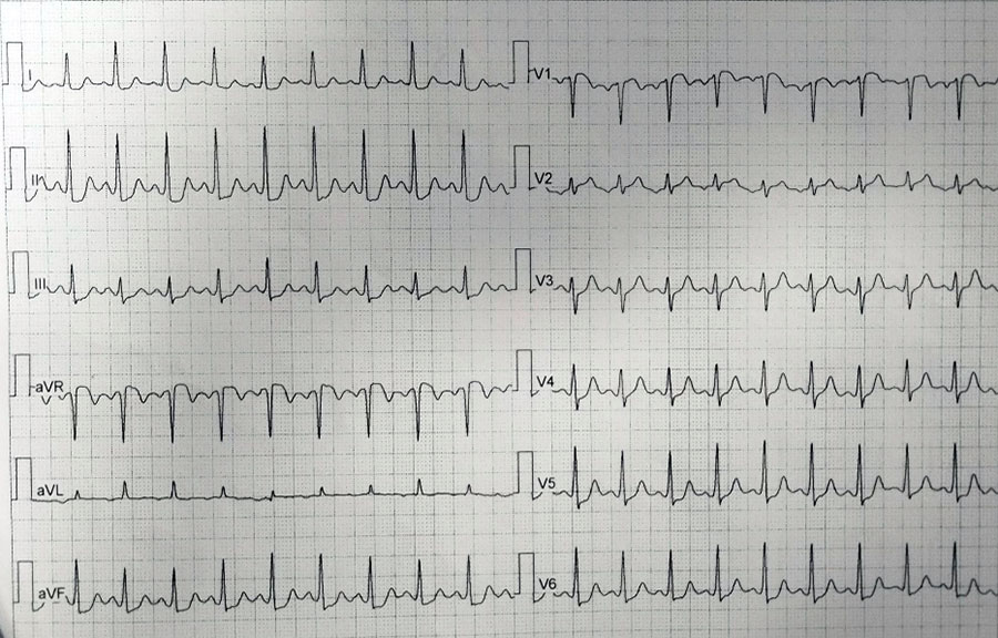
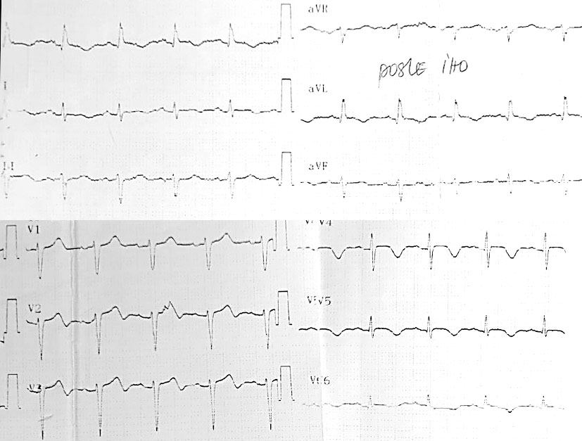
ECG at the admission:


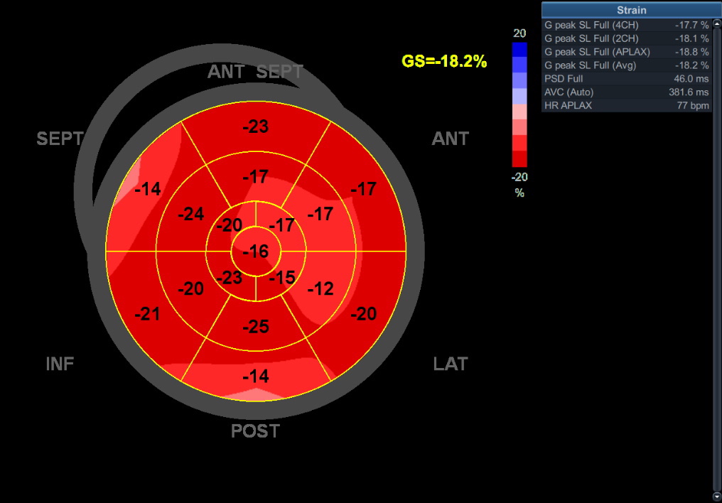
ECHO:
Normal dimensions of left and right heart chambers, LVEF estimated to be 50% with akinetic apical segments of the interventricular septum and inferior wall
Intrahospital course:
- Upon admission she was treated with Clopidogrel 75 mg o.d, Telmisartan 40 mg o.d., Rosuvastatin 20 mg o.d., LMWH (
- Laboratory analyses: Troponin I 95…292 ng/l (upper limit 0.05 ng/l) Creatin kinase 96…91 U/l NTproBNP 266 pg/ml total cholesterol 3.55 mmol/l LDL 1.52 mmol/l HDL 1.62 mmol/l triglycerides 0.90 mmol/l and all other taken were within normal range
CORONAROGRAPHY 2002.:
SCAD on LAD… images not available, upon medical documentation, presented as STEMI in August 2002.y, treated with thrombolytic therapy, successfully recovered, coronagraphy performed after one month, SCAD of the distal LAD described.
CORONAROGRAPHY 23.nov 2011.:
SCAD on OM 2
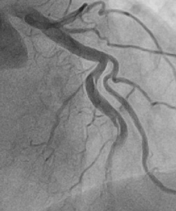
CORONAROGRAPHY 26.dec 2024.:
SCAD on D2
CORONAROGRAPHY 05.Jan 2025.:
No LMWH, on DAPT… no resolution, patient stable, without ongoing ischemia, due to the size of the artery intravascular imaging wasn’t performed
Instead of the conclusion:
- Dicharged home with DAPT (Aspirin 100 mg o.d., Clopidogrel 75 mg o.d., Pantoprazole 20 mg o.d., Bisoprolol 1.25 mg o.d., Valsartan 80 mg o.d., Amlodipine 5 mg o.d., Rosuvastatin 5 mg o.d., Folic acid 5 mg o.d. (during winter upon the recommendation of hematologist)
- MSCT angiography of the endocranium performed on 21.Jan 2025.y revealed a.cerebri media aneurysm
- Further diagnostic towards the underlying condition of the recurrent SCAD ongoing…
Author: Aleksandra Djokovic, MD, PhD, FESC, University of Belgrade, Faculty of Medicine, University Hospital Center Bezanijska kosa, Belgrade, Serbia



 ECG during chest pain:


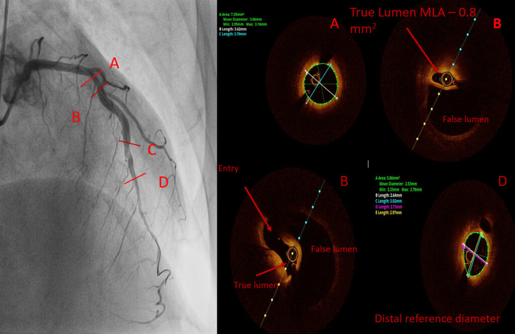
 DES 3.50×30 mm @ 12atm.
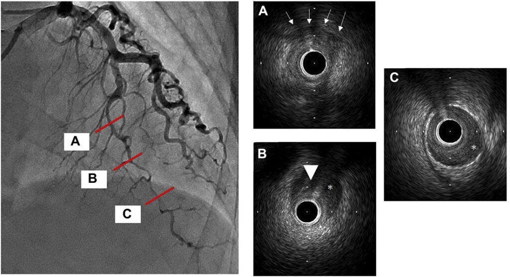
 After initial stabilisation patient was transferred to the tertiary center for coronary angiography and further diagnostic.


 Risk factors for CVD: hypertension, obesity (BMI 36), positive family history of CVD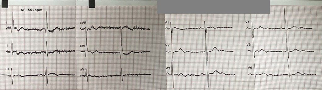


 LAB: TnI 2.180; K 3.1; Hol 4.47; LDL 2.49; HDL 1.24; TRIG 1.6
 ECHO day one:
Great images of the case! Since patient was without chest pain, with a TIMI 2-3 flow in LAD, would opt for conservative treatment (no PCI, no DAPT or LMWH, control angio before dismission). Best regards for colleagues from IKVB Sremska Kamenica!
It is a really great case. I am not an interventional cardiologist, however, it is very illustrative. And what did you do? Could you inform us about the FU of this patient, please?
Thank you for these experience.
I would also stay to a conservative approach.
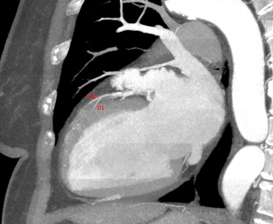
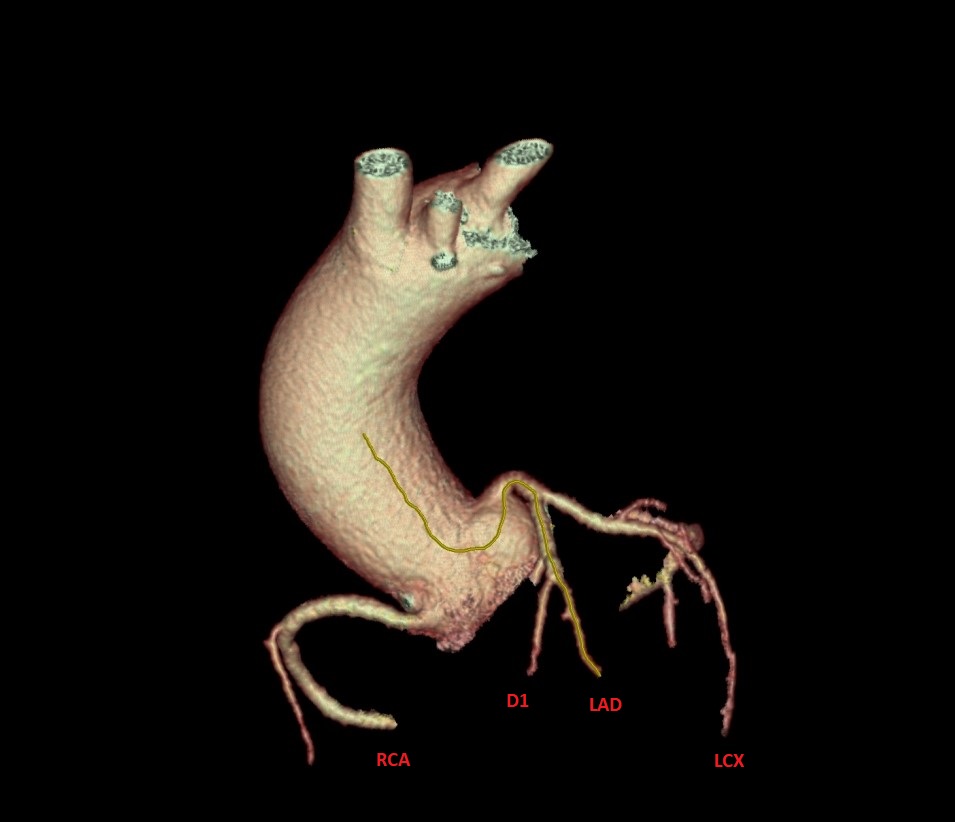
How do you explain the association of aortic dissection and SCAD?
What do you think about contrast retension before LM? Calcium?
Very interesting case. Of curse conservative approach is correct choice for this patients. Fibromuscular dysplasia can be pathophysiological mechanism who can explain aortic dissection and SCAD. Shadow (contrast retention) before LM is similar like calcium, but must think about aortic dissection especially in patients with history for aortic dissections and SCAD!
Since the patient had NSTEMI complicated with VF and OHCA I wouldn’t be brave enough to proceed with conservative treatment without a further diagnostic.
I would do intracoronary imaging to assess the distribution and MLA of the true lumen. If the true lumen is severely compromised I would proceed with PCI. I agree with Dr Mitov that FMD can be the cause.